
Shyama Nandakumar, Graduate Student, Department of Molecular Cellular and Developmental Biology (Buttitta Laboratory), University of Michigan
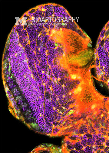
This is an image of the brain of a developing fruit fly larva. This lobe of the brain will become one optic lobe (connected to the fly eye) as well as half of the central brain. Purple marks the nuclei of all cells, and orange/gold marks all the membranes. The green marker shows proliferating stem cells in the brain. On the left-hand side of the image, you can see several layers of cells that are undergoing division in a stratified manner, producing the cell columns seen here. All the reporters except the DNA are encoded in the fly’s genes, so the flies just come out letting us know if their cells are dividing. We use this to study how the patterns of cell division in the brain change over time during development and aging.
18-016